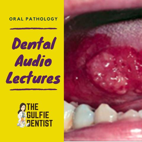
7. Mucosal autoimmune

Download and listen anywhere
Download your favorite episodes and enjoy them, wherever you are! Sign up or log in now to access offline listening.
Description
MUCOSAL – IMMUNOLOGIC These conditions are related to autoimmune (when our immune system attacks our own body) or hyperimmune (immune system just over reacts) reactions to some stimuli. Clinical manifestations...
show moreThese conditions are related to autoimmune (when our immune system attacks our own body) or hyperimmune (immune system just over reacts) reactions to some stimuli. Clinical manifestations include vesicles or bullae, ulcers, erythema, and white patches
They are treated with steroids !!
APHTHOUS ULCER
- Main cause Is stress
Site
- Present only in non keratinized tissue (opp to hsv infection)
- Ie; soft palate, buccal mucosa, ventral surface of tongue, labial mucosa,
Clinical Types of Aphthous Ulcers : MINOR & MAJOR
MINOR APHTHOUS ULCERS
- One to several painful oval ulcers 0.5 cm
- Very painful and may be debilitating
- May take several weeks to heal, even 21 days
- Will heal with scarring
- Rx Corticosteroids (triamcinolone ointment)
- Rx – Triamcinolone ointment or kenakort
Syndromes with Aphthous:
- Behcet syndrome – multiple aphthous ulcer + vasculitis
- Reiter syndrome – multiple aphthous ulcer + arthritis + urethritis + conjunctivitis
ERYTHEMA MULTIFORME
- Lesions seen on skin and mouth
- BULL’S EYE or Target or IRIS RIM lesion
- Allergic to medication like sulfa allergie, penicillin , barbiturate
- Infection like HSV + Mycoplasma
- Associated with Steven-Johnson Syndrome
- Type III hypersensitivity reaction
- Skin lesion + oral lesion + conjunctivitis + urethritis
- And Bull’s eye ulcers
- QN 🡪 the patient will have bulls eye on the skin and oral ulcers
LICHEN PLANUS
- Autoimmune disease of skin and mucous membrane
- Precipitating factors – stress + hep C***
- Site – skin + oral mucous membrane
- Variants – retricular lichen planus- most common
- Wickham’s striae
- H/F – civette bodies, rete pegs
- Grin’s span syndrome – hypertension + diabetes mellitus + lichen planus
- Rx – steroids (autoimmune na**)
- Long case picture shown white patches in buccal mucosa 15 yr old child had exams last week. Histopatholgy civatte bodies , hyperkeratosis etc
-Qn. Case 14 years old patient presents with white lace pattern lesions on skin and buccal mucosa, stressed, history of hep C 🡪lichen planus
- Rx corticosteroids
SYSTEMIC LUPUS ERYTHOMATOSIS
- Another auto immune disease
- Multiple organ involved
- Characteristic feature – BUTTERFLY RASH
- Rx corticosteroids
PEMPHIGUS VULGARIS
- Autoimmune – Ig G present
- Immune fluorescent test : +ve
- Most commonly affected site buccal 🡪 palatal 🡪lingual 🡪 labial
- Gingiva is least commonly affected site
- Auto antibodies against desmosomes**
- Rx – Steroids
C/F
- 1st Bullae + then painful vesicle
- Suprabasilar split**
- Acantholysis
- Intra epidermal
- Nickolskys sign +ve (also seen in Hailey – Hailey disease, toxic epidermolysis bullae)
Ie. When rubbing the affected skin 🡪 results in exfoliation of the skin
- Histopathology – Tzanck cells seen**
QN - The right corticosteroid daily dose for pemphigus vulgaris is: 50-100mg
STEROIDS - 100mg hydrocortisone. (Max. is 120mg. daily prednisone) 1-2 mg/kg/daily.
(max. is 120 mg. daily prednisone).
BULLOUS PEMPHIGOID
- Autoimmune – Ig G present
- Immune fluorescent test : +ve
- Auto antibodies against basement membrane**
- Rx – steroids
C/F
- Bullae + vesicle ( Remember it as BULLOUS PEMPHIGOID )
- Sub basilar split
- Sub epidermal bullae
- Nickolsky’s sign - -ve
- Desquamative gingivitis + skin lesion
Information
| Author | The Gulfie Dentist |
| Organization | The Gulfie Dentist |
| Website | - |
| Tags |
-
|
Copyright 2024 - Spreaker Inc. an iHeartMedia Company
